Get to know about Your Knee Replacement
Know Your Knee Replacement
- What is Knee Replacement?
- Who Is A Candidate For Knee Replacement?
- How Is The Knee Replaced?
- How is the recovery pattern at Naisarg Center?
- How Is The Artificial Implant Fixed To Bone?
- Frequently Asked Questions.
- Preparing For A Knee Replacement.
- Knee Implant Rates As Per NPPA (16_Aug_2017)
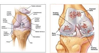
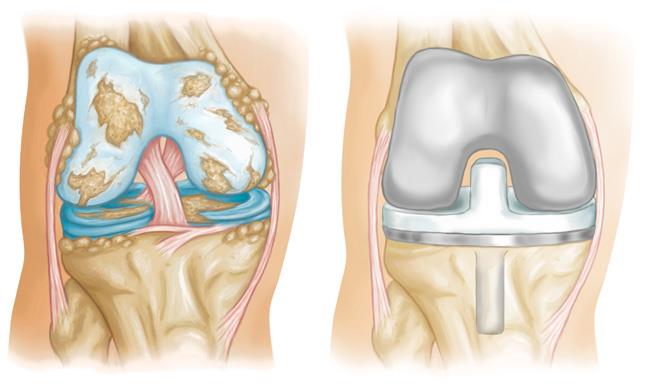
Normal Knee
Damaged Knee
What Is Knee Replacement?
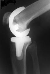
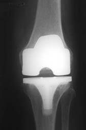
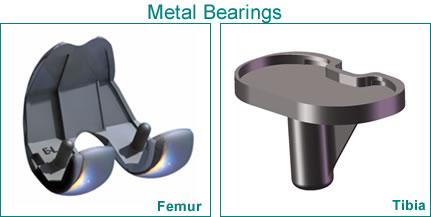
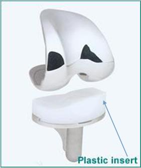
The total knee implant (TKR), also known as total knee arthroplasty, is a surgical treatment aimed at replacing the damaged or diseased parts of the patella bone areas with long-lasting and low-friction artificial components (materials). The prosthesis generally consists of two main substances: a metal part that is made up of titanium (Ti) or cobalt-chromium CoCr-based alloys for metal-on-metal joint replacement implants.
The Knee Implant
The primary objective of this safe and effective artificial structure implantation procedure is to restore pain-free natural movement and improve the quality of life, enabling independent standing, sitting, walking, and performing daily activities.
Nice to know:
Researchers indicate that over 95% of these implantations remain successful for at least 15-20 years if maintained properly. Moreover, fully functional joint replacement surgery in India was introduced in 1981.
Dr. Kalpit Patel from Naisarg Centre performed his first case in 2005, and since then he has been performing over 2000 successful TRK surgeries/operations.
Who Is A Candidate For Knee Replacement?
The eligibility for individuals having significant hinges issues and severe damage, adhering to meeting the below criteria:
- Those who have tried all non-surgical treatments with less success and relief,
- Patients experiencing severe lower limb hinge pain disrupting their normal activities in daily lives,
- The medically fit patients (in good mental and physical state), or above 50 years of age.
- X-rays are used to show the extent of damage to the joint, and they may suggest a cause for the degeneration.
X-rays are used to show the extent of damage to the joint and they may suggest a cause for the degeneration.


Additionally, blood tests or diagnostic tests like RA factor (rheumatoid factor), CBC (complete blood count), ESR (erythrocyte sedimentation rate), and serum uric acid may be required to identify infections like rheumatoid arthritis, bone infections, etc.
The lower portion (end) of the femur (distal femur) is covered with a metal cap, while the upper section of the tibia (proximal tibia) is resurfaced with the combined metal and plastic components fixed with screws or cement.
How Is The Knee Replaced?
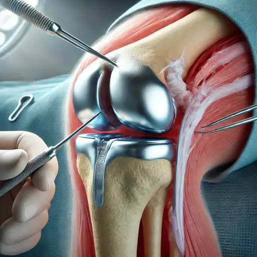
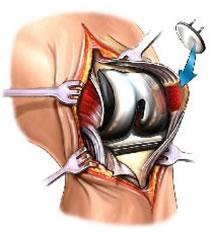
Once the anesthesia is administered, an incision of approximately 8 cm is made on the patella in front of the patient. Muscles and ligaments are carefully cut and preserved as much as possible. After this, the damaged hinge surfaces are removed from all three bones of the femoral-tibial area (thigh bone, shin bone, and patella cap), and the lower surface of the femur is capped with metal. The tibia’s upper end is fitted with a flexible metal and plastic implant rod, secured with cement and screws.
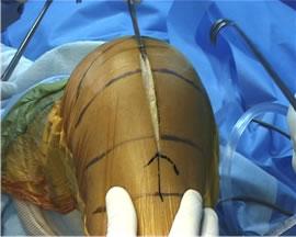
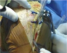
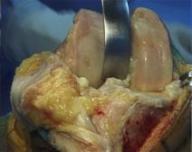
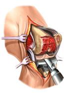
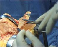
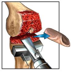
If arthritis infection does not affect the back of the kneecap (popliteal fossa), it can be preserved and left untouched. Highly specialized instruments are used that allow precision cutting of the bone so that the new structure fits perfectly. The surgeons consider all the major factors, like ligament stability, the patient’s age, overall health, femoral-tibial anatomy, etc., while selecting an appropriate type of patella implant for you.
Recovery Pattern in “NAISARG” Center For KNEE & HIP REPLACEMENT
On First day you can expect
We prefer spinal anesthesia, so you will be awake when you are brought back from Operation theater, but Oxygen (O2) will be given for approx. 2 hours for safety reasons.
We prefer spinal anesthesia, so you will be awake when you are brought back from the operating theater, but oxygen (O2) will be given for approx. 2 hours for safety reasons.
Water and liquids will be started gradually after 3 hours, and a light diet later on; if there is a feeling of nausea, antiemetics will be given. Blood transfusion is given if necessary.
The effects of spinal anesthesia and pain blocks usually subside within 4-6 hours, restoring the movements and sensations to the operated leg. After some time, gentle foot and joint exercises start with pain relief tablets to treat pain and infection. A negative suction draining tube is used to collect and remove the blood around the joint.
We prefer spinal anesthesia, so you will be awake when you are brought back from the operating theater, but oxygen (O2) will be given for approx. 2 hours for safety reasons.
You will be allowed to sit up and turn in bed and asked to start gentle foot and knee exercises.
Pain medication within safety limits are given by injection or as tablets There will be a tube draining collected blood around knee via negative suction.
On Second day you can expect
You can have regular meals and receive IV (intravenous) and anti-clotting medications after the wound drains are removed. You are instructed with safe movements and techniques.
You can use assistive devices like a walker, crutches, etc., for walking. Post-operative lab tests are conducted to examine the hemoglobin and kidney functions.
On Third day you can expect
You can expect wound dressing to be checked, and walking and femoral-tibial bending to be started by the physiotherapist. Switch over to oral medication from injectables.
On Fourth day you can expect
You can expect progression in walking and exercises; guidance and tips on safe bathroom use, instruction on dressing, advice on bed mobility, etc. are given to avoid stiffness, instability, and loss of function.
You can plan to go home with the necessary medicines and physiotherapy advice.
Advice On Discharge
“New Concept” – Self Physiotherapy
We are using the “Hi-Flex Joint” and advanced tissue release techniques to reduce the need for painful physiotherapy sessions and improve mobility.
Follow-up schedule :-
- Day 12-14: X-ray review and switches are removed.
- One Month: Independent walking, jumping, and stair climbing.
- Six Months: You can safely return to your normal daily routine.
How Is The Artificial Implant Fixed To Bone?
The artificial knee implant (man-made tailored components) is firmly attached to the bone with an adhesive called bone cement (polymethyl methacrylate (PMMA). This biomaterial is prepared by a chemical reaction between a special type of polymer and a liquid reagent.

Preparing For A Knee Replacement
Assessing the Fitness “ALL CLEAR” to undergo Surgery
- Blood /Urine test – A complete blood count, Liver and kidney function tests, blood sugar testing and any further test as advised by our physician depending on clinical need are done prior to surgery. Because there may be a need for blood transfusion during or after the surgery, blood tests will be needed for blood matching.
- ECG/ ECHO- Provides information regarding the condition of the heart for surgery
- Chest x-ray – Provides information about the respiratory status of the individual.
Eating and drinking instructions – you will be asked to stop all food and liquids 6 hours before surgery time. Fluids will be given through
IV line & Medications – The physician, anesthesiologist and nursing staff will need a current list of all prescription and non-prescription medications being taken by you. A sleeping tablet is given the night before surgery. A pre-operative antibiotic dose will be administered to fight against urinary and dental micro-organisms.
Our Success Stories

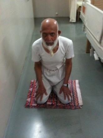
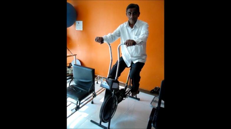
Cemented implants are stable prosthetic options offering immediate mobility. This is the most reliable, tried, and tested method that is best suited for people having poor bone quality. However, the downside of this is that after 15 years, it might require revision due to loosening.
